Weight loss may seem like a win-win pursuit. However, as naturopathic physicians and functional medicine providers, we need to be acutely aware of the risks associated with mobilizing toxins that have bio-accumulated over the months and years in adipose tissue.
Weight Loss Paradox- The Safety Question
An abundance of evidence indicates the obesity paradox can be explained by the release of toxins that occurs during weight loss. Adipose tissue is a known reservoir for toxins, and ironically, these toxins may have contributed to weight gain in the first place. However, the storage of persistent organic pollutants (POPs) in adipose tissue can prevent its build-up in other organs, therefore protecting against the otherwise toxic effects of POPs.
In a study of 63 participants, researchers collected serum samples before and one year after bariatric surgery. The subjects lost a mean of 70 pounds over a year. The study authors measured levels of POPs in the samples and found that after weight loss, there was a 46.7% to 83.1% increase in serum POPs levels.
There is evidence that not all fat is created equal in releasing toxins. One study found that the most significant release of toxins occurred in people who lost visceral adipose tissue compared to subcutaneous fat. This effect was only observed in the patients undergoing dieting and not those who lost weight through bariatric surgery.
Yet, the toxins sequestered in fat tissue are not completely harmless as they can exert inflammatory changes in the adipose tissue and be slowly released into the bloodstream over time.
Organic Acid Testing with Environmental Pollutants Panel
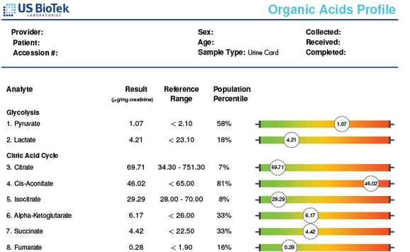
The best way to identify the presence of toxins in patients before undergoing weight loss is to order an organic acid profile with an environmental pollutants panel. This quantitative measurement of 14 select metabolites can help define individual toxic burdens. The test detects the presence of POPs such as xylene, toluene, benzene, trimethylbenzene, styrene, and chemicals like phthalates, parabens, and methyl tert-butyl ether. This test allows us as clinicians to better support in a focused manner the detox of pollutants that could be released during weight loss.
This panel's organic acid profile component helps identify the ability to utilize proteins, carbohydrates optimally, and lipids metabolism. The OAP panel also helps identify co-factors that may be suboptimal for well-fueled ATP production. Click for more information about our Organic Acids Profile.
Supporting Detox Pathways
Supporting detoxification pathways is critical to preventing unduly harmful mobilization of toxins during weight loss. In these detoxification pathways, fat-soluble toxins are converted into water-soluble substances for easier removal from the body. Identifying the specific toxins present in your patient allows for a customized approach to mitigate toxin release with successful weight loss.
Eating the Right Food While Dieting
Russian researchers diagnosed food intolerance to 13 food products in 32.6 to 33.4% of obese patients. According to the researchers, “The changes in immune status in obese patients created conditions for the development of food intolerance. The timely diagnosis of food intolerance allows to personalize the diet therapy.”
It is helpful to recognize that patients are immunologically unique beings. My approach is to test a patient for food intolerances/sensitivities before I change their diet, and ideally with a broad test of 208 foods and spices. I have found spices such as ginger, turmeric, and oregano are grand saboteurs that are often overlooked because they are used in small quantities unless they are used therapeutically. By performing food sensitivity/intolerance testing for IgG- and IgA-mediated reactions along with IgE allergy testing, patients can make empowered choices.
Conclusion
Certain steps should be taken before implementing a weight loss program, especially identifying the extent of the toxic burden and then recommending detoxification strategies. Performing an Organic Acid Profile/Environmental Pollutant panel along with food sensitivity/intolerance testing prior to undertaking a weight loss protocol is vital to ensure patients are not eating foods that are sabotaging their health.
Watch Our Webinar:
Register for our upcoming webinar: Weight Lose 101: Mitigating the Risk of Liberating ‘Fat Stored Toxin’ to explore how losing weight without correctly identifying a patient’s toxic burden and individualizing their detox protocol may indeed put them at risk.
References:
Lee DH. Persistent organic pollutants in adipose tissue should be considered in obesity research. Obes Rev. 2017;18(2):129-139.
Hong NS, Kim KS, Lee IK, et al. The association between obesity and mortality in the elderly differs by serum concentrations of persistent organic pollutants: a possible explanation for the obesity paradox. Int J Obes (Lond). 2012;36(9):1170-1175.
Jansen A, Polder A, Müller MHB, Skjerve E, Aaseth J, Lyche JL. Increased levels of persistent organic pollutants in serum one year after a great weight loss in humans: Are the levels exceeding health-based guideline values? Sci Total Environ. 2018;622-623:1317-1326.
Dirinck E, Dirtu AC, Jorens PG, Malarvannan G, Covaci A, Van Gaal LF. Pivotal Role for the Visceral Fat Compartment in the Release of Persistent Organic Pollutants During Weight Loss. J Clin Endocrinol Metab. 2015;100(12):4463-4471.
La Merrill M, Emond C, Kim MJ, et al. Toxicological function of adipose tissue: focus on persistent organic pollutants. Environ Health Perspect. 2013;121(2):162-169.
Sentsova TB, Gapparova KM, Grigor'ian ON, Vorozhko IV, Kirillova OO, Chekhonina Iu G. [The clinical and immunological manifestations of food intolerance in obese patients]. Vopr Pitan. 2012;81(5):83-87.
Cioffi F, Senese R, Lanni A, Goglia F. Thyroid hormones and mitochondria: with a brief look at derivatives and analogues. Mol Cell Endocrinol. 2013;379(1-2):51-61.

